Trametes aesculi
Scientific name: Trametes aesculi (Fr.) Justo
Derivation of name: Trametes means "one who is thin;"
aescul- means "oak."
Synonymy: Daedalea elegans Spreng.: Fr.; Daedalea
ambigua Berk.; Lenzites elegans (Spreng.) Pat.; Trametes
elegans (Spreng.:Fr.) Fr.
Common names: Unknown
Phylum: Basidiomycota
Order: Polyporales
Family: Polyporaceae
Occurrence on wood substrate: Saprobic; scattered to
grouped, often overlapping on decaying deciduous wood;
July through December.
Dimensions: Caps 2-14 cm wide; stipes absent or
rudimentary.
Upper surface: Whitish to grayish or pale ochraceous or
even blackish toward the base in older specimens;
greenish from the base outward when covered by algae;
glabrous at maturity; warted or not; often
concentrically grooved (sulcate).
Pore surface: Whitish, becoming pale ochraceous in age;
variable in that within the same specimen parts of the pore
surface are poroid, gill-like, and maze-like; pores 1-2 per
mm.
Edibility: Inedible.
Comments: This species has often been recorded as
Trametes elegans during forays but Trametes elegans
apparently does not occur in the United States or
Canada. Trametes gibbosa is very similar but has a
conspicuosly uneven warty or bumpy upper surface,
hence the common name "lumpy bracket." The upper
surface of T. gibbosa is also described as distinctly
velvety or fuzzy when observed with a hand lens.
Unfortunately, there is enough overlap in
macroscopic characters that confident identification
may not be possible without measuring spores. In fact,
because the photographs on this page were obtained
prior to my awareness of the confusion
of species, I
am not certain that every photograph is of T. aesculi.
More information at MushroomExpert.com:
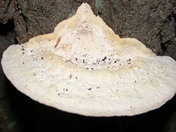
Figure 1. Upper surface of Trametes aesculi.
Photo ©
Tom Volk.
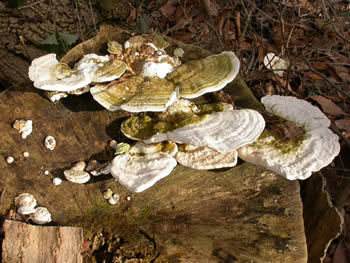
Figure 2. The fruit bodies are persistent. This photo
was taken in late December.
Photo © Gary Emberger.
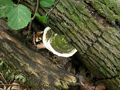
Figure 3. The greenish coloration at the base of this old
specimen is due to the growth of algae.
Photo
© Diana Smith.
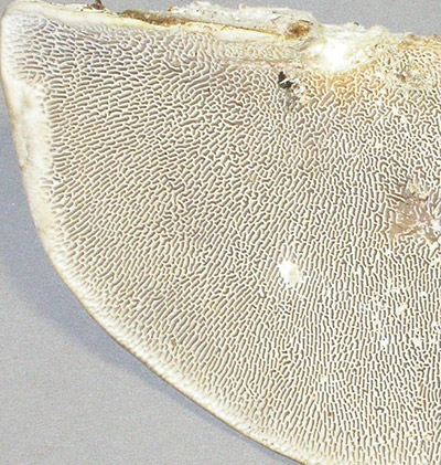
Figure 4. Variation in pore surface from cap margin
to
the point of attachment (at top).
Photo © Gary Emberger.
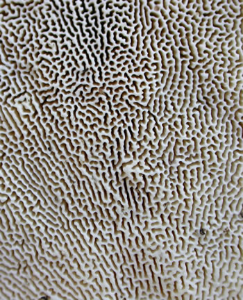
FIgure 5. Maze-like area of pore surface of Trametes
aesculi. Photo © Gary Emberger.
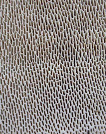
Figure 6. Poroid area of the fertile
surface
of Trametes
aesculi. Note the radial elongation of the pores.
Photo © Gary Emberger.
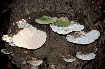
Figure 7. One last look at Trametes aesculi growing on the cut
end of a tree trunk.
Photo © Larry Grand.